科技工作者之家
科技工作者之家APP是专注科技人才,知识分享与人才交流的服务平台。
科技工作者之家 2019-05-16
来源:研之成理
▲共同第一作者:郭兵,陈静钦,陈宁波;通讯作者:刘成波,刘斌;
通讯单位:新加坡国立大学,深圳先进技术研究院;
论文DOI:10.1002/adma.201808355
全文速览
体外造影剂辅助的近红外二区光声显微成像技术可以解析三维广面积/大深度,高信号/背景比例,高成像深度/深度分辨率比例的生物组织。本文章首次报道了就具有强近红外二区吸光系数的共轭高分子纳米颗粒辅助的三维高分辨率光声显微成像,实现了脑部和肿瘤部位的肿瘤心血管的微米分辨率,毫米穿透深度,高信号/背景比例的原位活体成像。此文成像效果比近红外二区荧光三维激光共聚焦成像效果更有优势。
背景介绍
生物组织三维成像技术可以用来解析血管和肿瘤结构,有利于分析生理/病理过程,是目前成像技术发展的前沿。传统三维成像技术各自有一定局限性。比如核磁, PET 和 CT 成像分辨率不足。双光子和近红外二区荧光激光共聚焦成像的视野狭小,其成像质量有待进一步提高。光声显微成像能够调节分辨率和成像深度,是近年来新兴的成像技术。相对于近红外一区/可见光光声显微成像,近红外二区光声显微成像能够就降低光散射/生物组织光吸收对成像的干扰。此前报道的近红外二区光声成像大都使用体内造影剂来成像, 但是生物组织容易产生强噪音干扰,使体内造影剂辅助近红外二区光声成像表现出低信号/背景比例,模糊的成像效果。
研究出发点
体外造影剂能够显著提高成像质量. 适用于活体成像的造影剂需要生物相容性好,光稳定性好,吸光系数大,弱荧光,可大规模制备等特点. 有机共轭高分子可以满足这些条件. 于是, 我们设计了一个新型, 微流控技术制备的具有强近红外二区吸收的共轭高分子纳米颗粒, 来辅助实现三维近红外二区显微光声成像。
图文解析
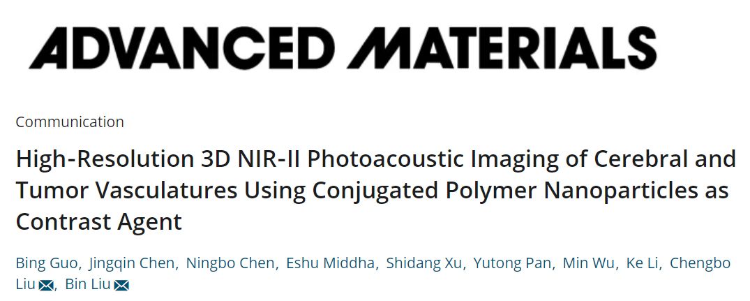
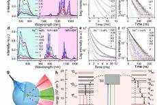
刘斌课题组设计了新型电子给体-电子受体1 -电子给体-电子受体2 结构构成的共轭高分子 PTD, 使用课题组自制的微流控技术制备了大小可控且尺寸均一的纳米颗粒(40 纳米左右). 该纳米颗粒在吸收峰 1161 纳米左右的吸光系数 高达 48.1 L g-1, 有利于实现高效光声造影 (如图1 所示)。
▲Figure 1. (a) The synthetic route towards PTD. Reagents and conditions: Pd2(dba)3, P(o-tyl)3, anhydrous toluene, 100 oC, 48 h; (b) Schematic diagram of microfluidic glass capillary mixer for the synthesis of monodisperse PTD NPs through modified nanoprecipitation. (c) TEM image of PTD NPs synthesized at Re 320 with 40% EtOH in the anti-solvent. (d) Changes in the size and PDI of PTD NPs by varying the amount of EtOH in the antisolvent from 0 to 75% at Re 320. (e) Variation in the size of PTD NPs with 25, 40 and 75% EtOH in the anti-solvent at different Re.
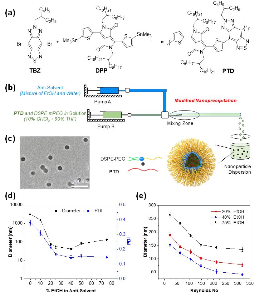
同时, 如图2 所示, 我们使用该纳米颗粒实现了耳朵上皮下肝肿瘤的血管三维成像. 首先, 在未注释纳米颗粒前,调整光声成像参数,使背景信号降低到最低. 注射纳米颗粒后,使用 1064 纳米脉冲激光, 实现了肿瘤部位的无损广面积成像. 成像面积高达 7 毫米× 7 毫米, 成像深度达 0.76 毫米. 在 755 微米成像深度处, 分辨率是 25.9 微米, 信号/背景比例是 26.0 dB, 成像深度/深度分辨率高达 29.1 倍. 同时通过定性和定量比对肿瘤部位和周围正常组织的血管密度, 肿瘤边界可以勾画出来. 本研究的近红外二区光声显微成像效果比最近报道的近红外二区荧光三维共聚焦肿瘤成像效果好.可能原因有三个: (1) 本实验使用的 1064 纳米激发光比近红外二区荧光成像使用的808纳米激光的光散射效应更低; (2) 光声成像的声波散射比荧光成像的光散射效应更低; (3) PTD 纳米颗粒吸光系数大, 可以最大限度的抑制背景噪音. 然而二区荧光中的活体自发荧光比较强, 产生的噪音干扰不可忽略。
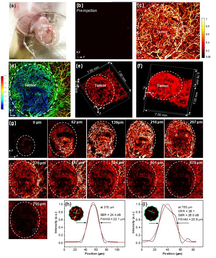
此外, 如图3 所示, 我们使用 PTD 纳米颗粒实现了透过老鼠头骨脑血管的三维高分辨(分辨率 25.4 微米),高信号/背景比例( 22.3 dB)成像. 其成像深度高达 1001 微米. 该脑血管光声成像效果比最近报道的双光子成像和近红外二区荧光共聚焦成像的效果好.
▲Figure 2. PA imaging of subcutaneous HepG2 tumor-bearing mouse ear with a colorbar 0.06-1. (a) Photo of mouse ear bearing subcutaneous tumor for PA imaging. Representative xy projected tumor bearing mouse ear image (7.00 × 7.00 mm, x × y) before (b) and after (c) PTD NP administration. (d) Depth-encoded maximum amplitude projection image corresponding to Figure c (The PA signal color changes correspond to different depths according to the color chart for depth information on the right side). (e) and (f) 3D reconstruction of tumor-bearing mouse ear vasculature images from different view side (7.00 × 7.00 × 0.76 mm, x × y × z) and the tumor margin was labelled with white-dashed circle. (g) Layer-by-layer PA images (7.00 × 7.00 mm, x × y) of subcutaneous tumor-bearing mouse ear with white-dashed circle for labelling tumor margin in each layer. (h) and (i) The PA intensity profile (black curve) along the green line in the zoomed area (insets, Figures h and i) which represents the area labelled with green-dashed circle at depths of 370 and 755 µm, respectively. The Gaussian fits to the profiles are presented using red curves. Gaussian-fitted full width at half maximum (FWHM) of the vessel along the green line is presented at different depth.
▲Figure 3. In vivo ORPAMI of whole-cortex brain through intact skull after administration of PTD NPs through tail-vain (colorbar: 0.06-1). (a) Layer-by-layer PA images (8 × 6 mm, x × y) of mouse brain. The deepest area reached 1001 µm. (b) Photo of mouse for imaging. (c) Representative xz projected brain vasculature image (8 × 1 mm, x × z). (e) Representative xy projected brain vasculature image (8 × 6 mm, x × y). (f) 3D reconstruction of brain vasculature (8 × 6 × 1 mm, x × y × z). (d) and (g) The PA intensity profiles along the green line in the zoomed area (inset, Figure d and g) which represents the area labelled with green-dashed circle (Figure a) at the depths of 77 and 1001 µm, respectively. The Gaussian fits to the profile are shown in red curve. Gaussian-fitted full width at half maximum (FWHM) of the vessel along the green line is presented at different depth.
总结与展望
我们首次实现了体外造影剂辅助近红外二区光声显微成像. 微流控技术制备共轭高分子, 可以实现尺寸可控, 形貌均一. 同时,共轭高分子生物相容性好,吸光系数大,光声稳定性好,是很好的活体成像的光声造影剂. 我们证明二区共轭高分子辅助光声显微成像可以准确勾画肿瘤边界, 解析肿瘤内部和周围正常组织血管结构, 准确成像脑补三维复杂血管脉络. 因此, 共轭高分子纳米颗粒是很有潜力的活体成像造影剂, 用来理解生理和病理过程。
心得与体会
这个工作的灵感来自于近红外二区荧光脑血管成像, 也是我们团队的光声显微成像的第一个工作. 刘成波老师团队搭建光声成像系统后, 刘斌老师团队筛选材料,设计了一种强吸收共轭高分子材料. 这篇文章充分体现了合作的重要性,合作双方取长补短,才能取得最好的科研成果.
课题组介绍
刘斌课题组致力于共轭聚合物发光材料、聚集诱导发光材料等在生物医学及能源中的应用研究,其成果多次发表在国际一流期刊。刘斌教授连续多年荣获科睿唯安“高被引科学家”称号。
来源:rationalscience 研之成理
原文链接:http://mp.weixin.qq.com/s?__biz=MzIwMzE5MzQ1NQ==&mid=2649325701&idx=3&sn=3e3f299a5ff75751205f600d185fb0a4&chksm=8ece0d85b9b98493a4cc281b58ef75aef005519ad30df0d1e67f20cf26ca605d872ba605a9ea&scene=27#wechat_redirect
版权声明:除非特别注明,本站所载内容来源于互联网、微信公众号等公开渠道,不代表本站观点,仅供参考、交流、公益传播之目的。转载的稿件版权归原作者或机构所有,如有侵权,请联系删除。
电话:(010)86409582
邮箱:kejie@scimall.org.cn
研究揭示二维荧光功能金属有机框架材料纳米片在水中离子检测传感的应用优势
范楼珍教授课题组在荧光碳纳米材料领域取得重大突破

福建物构所荧光温度探针材料研究获进展
纳米级厚度氧化铝涂层或可完美防腐

蓝光LED可激发的近红外二区稀土掺杂CaS纳米荧光标记材料

中美研究人员开发出三维纳米“剪纸”结构

合工大研发智能水凝胶,一分钟实现96%的自修复
福建物构所研制出蓝光LED可激发的 近红外二区稀土掺杂CaS纳米荧光标记材料


新型超分辨显微镜首次将DNA纳米结构形象化

每日材料前沿|福建物构所热响应荧光材料研究取得进展


科技工作者之家APP是专注科技人才,知识分享与人才交流的服务平台。
 微信
微信
 京公网安备11010202008424号