科技工作者之家
科技工作者之家APP是专注科技人才,知识分享与人才交流的服务平台。
科技工作者之家 2019-06-04
来源:科研圈
俄勒冈大学领导的一项研究分析比较了帕金森患者和健康人的未经过滤的 β 脑波,发现了帕金森病标志物的新线索。
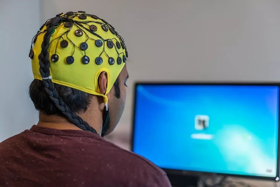
图片来源:Pixabay
来源 University of Oregon
翻译 页一
审校 刘悦晨
编辑 戚译引
β 波是人的四种基本脑波之一。研究人员在分析头皮脑电图(scalp electroencephalograms)未经过滤的原始数据时发现,β 脑波呈现的特定角度和尖锐程度可能与帕金森病有关。
该研究发表于网络期刊《神经在线》(eNeuro)。研究人员提出,非侵入性脑电图的读数可能提供了易于检测的电生理生物标记。这不仅将有助于帕金森病的诊断和精准治疗,而且可能同样适用于其它的运动障碍病症。
“通过这个安全且经济的方法来测量和量化大脑活动,我们能够区分出帕金森患者在用药和不用药时的差别,并可以同健康人进行比较,”这项研究的首席研究员,俄勒冈大学(University of Oregon)人类生理学系的妮可·斯万(Nicole Swann)说道。
目前,帕金森病的初次诊断需要患者完成一系列身体运动任务,让神经科医生进行评估。但是,这种主观诊断不可避免地存在误诊风险。
“我们目前还不知道这种新的诊断方法是否更好,但它可以提供容易获得的大脑测量数据,这可能对诊断有所帮助,还有可能与临床观察数据及其它的脑电图读数一起使用,”斯万说。
斯万说,在此之前用脑电图检测帕金森病的研究方法与这一新方法有所不同,因为此前的关注重点一直被放在将大脑信号建模为正弦波的方法上,即对 β 波进行过滤,以调整波形,并显示周期性振荡的总体强度。然而,类似本研究提出的方法此前需要依靠手术植入大脑的电极,才能发现 β 波的变化与帕金森病的关联。
本次研究的数据来自加州大学圣地亚哥分校(University of California San Diego),包含过去收集的 15 名帕金森患者和 16 名健康的对照组参与者的头皮脑电图数据,研究对数据进行了新的分析。斯万把重点放在了原始的未经过滤的 β 波上,寻找振动中特殊的角度和完整的波形。论文的共同作者斯科特·科尔(Scott Cole)是加州大学圣地亚哥分校 2017 年的一名博士生,他注意到帕金森氏症患者似乎有尖锐的脑电波,这表明他们有必要重新审视这些数据。
斯万说:“原始脑电波信号像正弦波一样上下振动,但不对称程度更高。波形的陡峭程度,也就是波形的斜率似乎对帕金森患者的病情诊断而言很重要。这在停止用药的患者身上很容易发现。”
她说,当波峰顶点比波谷(振荡下凹部分的)顶点更尖锐时,这表明患者可能已经停止用药。
斯万表示,她希望进行一项大规模的随访研究,将头皮脑电图的测量结果与详细的病史和患者每天测试时的自我感觉报告结合起来。最终,这种方法有望帮助我们检测个体病情随时间的变化。
本研究的共同作者布拉德利·沃泰克(Bradley Voytek)说,目前对帕金森病的治疗方法包括手术永久植入电刺激器或药物治疗,这两种方法的剂量都很难控制,往往导致令人沮丧的治疗效果循环。沃泰克是加州大学圣地亚哥分校的神经科学家,科尔获得博士学位之后曾在他的实验室工作。
沃泰克说:“如果能够实时衡量治疗能够在多大程度上减轻帕金森病症状,那么治疗剂量就可以实时调整。在使用侵入性脑刺激器的情况下,这可能意味着只在需要的时候使用电刺激。在使用药物治疗的情况下,这意味着调整药物的剂量——就像持续监测血糖的植入型血糖仪,它可以向泵发出信号,根据需要调整胰岛素水平。”
斯万说,理想情况下,如果这种方法提供的信息被证明可靠,那么帕金森患者就可以远程进行脑电图检测;他们只需戴着装有电极的帽子,然后将结果发送给他们的神经科医生,就能快速便捷地调整治疗。
但沃泰克指出,该设想面临的一个挑战是大脑活动的实时测量数据往往是充满噪声的,这会影响获取正确信号的能力。
他说:“我们现有的很多测量大脑活动的工具都依赖强大的数据处理能力,所以不容易应用于实时监测。”
论文信息
【标题】Characteristics of Waveform Shape in Parkinson’s Disease Detected with Scalp Electroencephalography
【作者】Nicko Jackson, Scott R. Cole, Bradley Voytek and Nicole C. Swann
【时间】2019 年 5 月 20 日
【期刊】eNeuro
【DOI】10.1523/ENEURO.0151-19.2019
【链接】http://www.eneuro.org/content/early/2019/05/10/ENEURO.0151-19.2019
【摘要】Neural activity in the beta frequency range (13-30 Hz) is excessively synchronized in Parkinson’s Disease (PD). Previous work using invasive intracranial recordings and non-invasive scalp electroencephalography (EEG) has shown that correlations between beta phase and broad-band gamma amplitude (i.e., phase amplitude coupling) are elevated in PD, perhaps a reflection of this synchrony. Recently, it has also been shown, in invasive human recordings, that nonsinusoidal features of beta oscillation shape also characterize PD. Here we show that these features of beta waveform shape also distinguish PD patients on and off medication using non-invasive recordings in a dataset of 15 PD patients with resting scalp EEG. Specifically, beta oscillations over sensorimotor electrodes in PD patients off medication had greater sharpness asymmetry and steepness asymmetry than on medication (sign rank, p<0.02, corrected). We also showed that beta oscillations over sensorimotor cortex most often had a canonical shape, and that using this prototypical shape as an inclusion-criteria increased the effect size of our findings. Together our findings suggest that novel ways of measuring beta synchrony that incorporate waveform shape could improve detection of PD pathophysiology in non-invasive recordings. Moreover, they motivate the consideration of waveform shape in future EEG studies.
Significance Statement Diagnosis and long-term monitoring of Parkinson's disease (PD) is mainly assessed via clinical rating scales which are subjective and can be imprecise. An objective measure of PD would be extremely valuable, especially one that could be acquired non-invasively. Here we show, using scalp electroencephalography (EEG), that the nonsinusoidal shape of beta (13-30 Hz) oscillations over sensorimotor cortex distinguishes PD patients on and off medication and patients off medication from controls. This change in waveform shape may reflect hypersynchronous input, possibly originating from basal ganglia. Thus, waveform shape is a putative non-invasive electrophysiology biomarker of PD state with potential utility for assessing treatments, monitoring disease, or diagnosis. This signature can be detected with a safe, affordable, and available method (i.e. EEG).
来源:keyanquan 科研圈
原文链接:http://mp.weixin.qq.com/s?__biz=MzA5NDkzNjIwMg==&mid=2651681233&idx=3&sn=87a4b6f988df3d2dc6c8760128941a87&chksm=8bbedc34bcc955221ffb8f1d33ee9994395378edceb49d3c758aa09b5e80e97d854f14dacc86&scene=27#wechat_redirect
版权声明:除非特别注明,本站所载内容来源于互联网、微信公众号等公开渠道,不代表本站观点,仅供参考、交流、公益传播之目的。转载的稿件版权归原作者或机构所有,如有侵权,请联系删除。
电话:(010)86409582
邮箱:kejie@scimall.org.cn

简单的眼部检查将可诊断帕金森病

世界帕金森病日:专家权威解读 科学防治帕金森病
新的帕金森病生物标志物可能就藏在脑电图里
脑电图可在婴儿3个月时高效预测自闭症

世界帕金森病日 | 如何正确认识帕金森病

日本批准用诱导多能干细胞治疗帕金森病
血液中的咖啡因浓度有望成为帕金森病诊断方法

世界帕金森病日 | 手抖,就一定是帕金森病吗?
多巴胺能提高帕金森患者“运动能量”

世界帕金森病日 | “帕金森”偏爱谁


科技工作者之家APP是专注科技人才,知识分享与人才交流的服务平台。
 微信
微信
 京公网安备11010202008424号